Stroke 101: What Everyone Needs to Know on World Stroke Day

October 27, 2023

Position title

What Is World Stroke Day?

World Stroke Day is an annual event observed on October 29th, which aims to raise awareness about stroke and its global impact. World Stroke Day is an annual event observed on October 29th, which aims to raise awareness about stroke and its global impact.

Organized by the World Stroke Organization (WSO), this day focuses on educating the public on the signs and symptoms of a stroke, the importance of immediate medical attention, and the long-term care and support that stroke survivors may need. The event also emphasizes the prevention strategies individuals can adopt to reduce their risk of a stroke.

This article seeks to demystify the basics of a stroke, recognizing early signs, preventing strokes through healthy lifestyle choices, and the science behind it, ensuring that you walk away with a clear understanding.

Understanding the Basics of a Stroke

What is a Stroke?

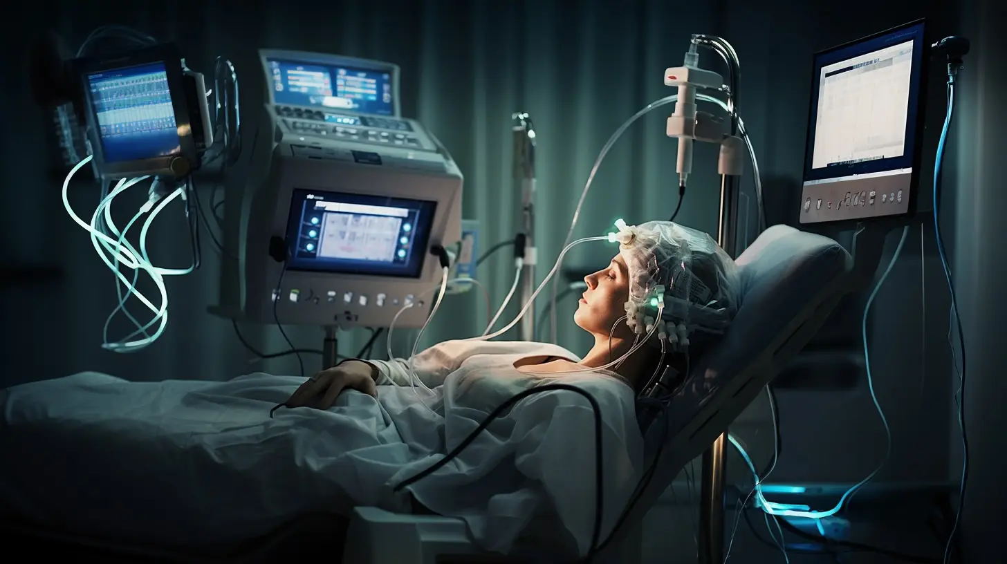
A stroke is a sudden interruption in the blood supply to the brain. Whether because of a blockage or bleeding, it’s a medical emergency that requires immediate attention. With millions affected by it globally, strokes are a leading cause of death and disability.

Why Do Strokes Happen?

At its core, a stroke is about the brain not getting the nutrients and oxygen it needs to function, typically due to blocked or ruptured blood vessels. Without the vital flow of blood, brain cells start to die, leading to potential long-term damage or death.

Different Types of Strokes
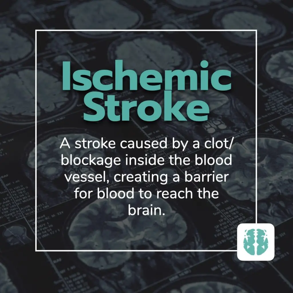
Ischemic Stroke

This type of stroke occurs when there’s a blockage in a blood vessel supplying blood to the brain. It’s the most common kind, accounting for about 80% of all strokes. These blockages are typically caused by blood clots or the buildup of fatty deposits and other debris.
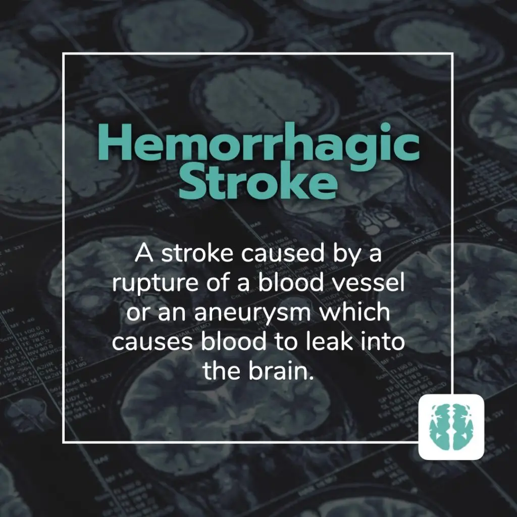
Hemorrhagic Stroke

Hemorrhagic strokes are caused by bleeding within or around the brain. This can result from conditions like aneurysms, which are weakened, bulging sections of blood vessel walls, or arteriovenous malformations (AVMs), tangles of malformed blood vessels.

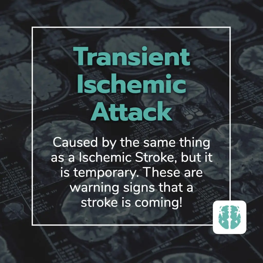
Transient Ischemic Attack (TIA)

Often referred to as a “mini-stroke”, a TIA resembles an ischemic stroke but doesn’t last as long and doesn’t cause permanent damage. Even though the effects of a TIA are temporary, it’s crucial to seek medical attention as it could be a warning sign of a future stroke.

The Science Behind a Stroke

Brain Cells and Their Need for Oxygen

Each of our brain cells needs a steady supply of oxygen to function. This oxygen is delivered via the blood. When there’s an interruption, brain cells become deprived and begin to die, which can lead to lasting damage or even death.

How Blood Flow Gets Interrupted

Picture a smoothly flowing river, and now imagine a dam suddenly blocking it. That’s what happens during an ischemic stroke. On the other hand, imagine the riverbank suddenly bursting, causing flooding—that’s akin to a hemorrhagic stroke.

Recognizing the Signs of a Stroke

The BEFAST Method:

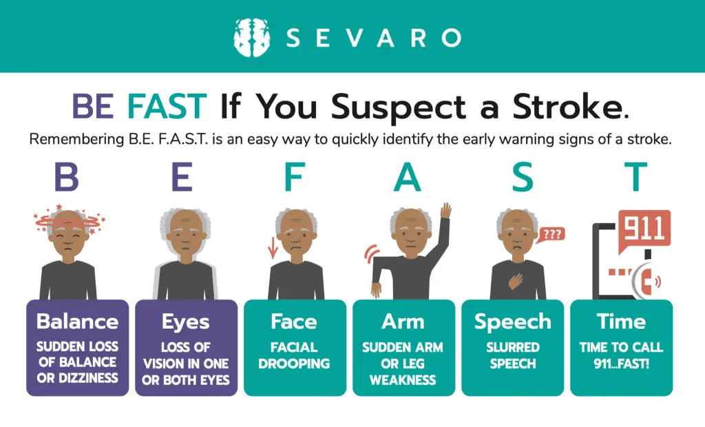
When suspecting someone might be having a stroke, remember the acronym BEFAST:

  • B (Balance): Sudden loss of balance or coordination.
  • E (Eyes): Sudden trouble seeing out of one or both eyes or double vision.
  • F (Face): Ask the person to smile and check if one side of the face droops.
  • A (Arms): Ask the person to raise both arms and see if one arm drifts downward.
  • S (Speech): Ask the person to repeat a simple phrase and listen for slurred or strange speech.
  • T (Time): If you notice any of these signs, it’s time to call 911 or emergency services immediately.

Other Potential Stroke Warning Signs

Keep an eye out for sudden numbness or weakness, especially on one side of the body, sudden confusion, trouble seeing in one or both eyes, trouble walking, dizziness, or an unexplained severe headache.

The Importance of Immediate Action For Stroke

Time is Brain

In stroke treatment, every second counts. The earlier a person gets treatment, the better their outcome is likely to be. In fact, getting to a hospital within 4.5 hours of the first symptoms can greatly reduce the risk of long-term disability.

Seek Immediate Medical Attention For Strokes

When you or someone else displays signs of a stroke, it’s essential to call emergency services right away. Also, remember to note when the symptoms first began, as this can influence treatment decisions.

Stroke Prevention: Lifestyle Choices That Make a Difference

What are the biggest Stroke Risk Factors?

  • High blood pressure: Often termed the “silent killer,” it’s the leading cause of stroke.
  • Diabetes: High sugar levels can lead to clotting more easily.
  • High cholesterol: Fatty deposits can clog your blood vessels.
  • Smoking: Nicotine raises your blood pressure.
  • Alcohol consumption: Excessive drinking can lead to multiple health complications, including stroke.
  • Family history: Stroke can sometimes run in the family.

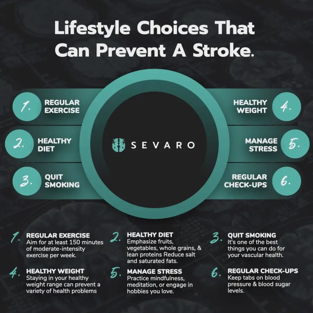
The Power of a Balanced Diet

Eating a balanced diet is vital for overall health and particularly beneficial for stroke prevention.

  • Incorporate whole grains, fruits, and vegetables: These provide essential nutrients and antioxidants.
  • Limit saturated fats and sugars: Helps maintain a healthy weight and reduces cholesterol.
  • Importance of hydration: Drinking water aids in maintaining blood volume and viscosity.

Embracing Regular Exercise

Physical activity has numerous health benefits, one of which is stroke prevention.

  • Aerobic activities: Walking, cycling, or swimming can improve heart health and reduce stroke risk.
  • Strength training and flexibility exercises: Builds muscle and maintains joint health.
  • The connection between physical activity and reduced stroke risk: Active individuals generally have lower cholesterol and healthier blood pressure levels.

The Importance of Smoking Cessation

Smoking accelerates clot formation by thickening your blood and increasing the amount of plaque buildup in arteries.

  • How smoking increases stroke risk: Nicotine and carbon monoxide from inhaling smoke reduce the amount of oxygen your blood can carry.
  • Benefits of quitting smoking: Enhanced lung capacity, reduced blood pressure, and improved blood circulation within a few weeks.

Regular Health Check-ups: Prevention is Better than Cure

It’s essential to keep an eye on potential risk factors.

  • Monitoring blood pressure: Consistent high readings are a significant red flag.
  • Regular cholesterol checks: Ensures you maintain a healthy balance of lipids.
  • Diabetes management: Monitoring sugar levels to prevent complications.

The Digital Revolution: How Technology is Transforming Stroke Care with telestroke

The Pivotal Role of Technology in Stroke Care

Historically, diagnosing and treating strokes depended heavily on a patient’s symptoms and basic imaging. Now, we harness technology to offer more precision, speed, and efficiency.
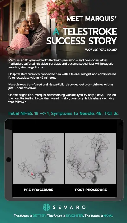
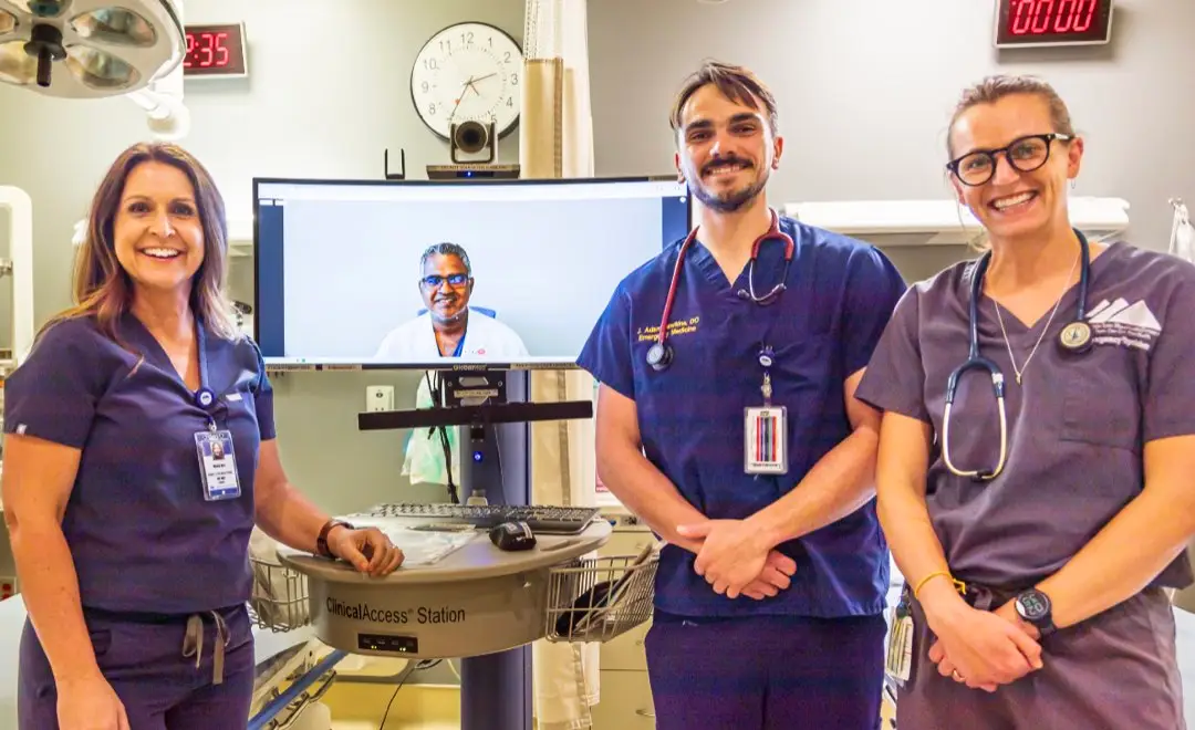
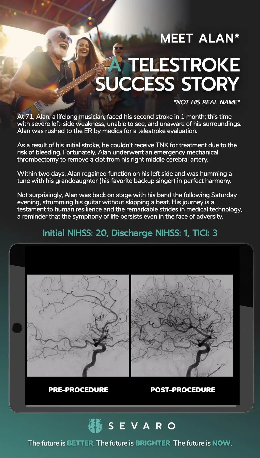
What is Telestroke?

Telestroke is a telemedicine approach that facilitates the consultation of stroke specialists for patients in remote or underserved areas using digital communication.

Benefits of Telestroke Services

  • Immediate access to specialists: Reduces the ‘door-to-needle’ time.
  • Reducing time-to-treatment: Prompt treatment can mean the difference between recovery and long-term disability.
  • Improved outcomes for patients: Access to specialists leads to better patient care and outcomes.

Revolutionizing Stroke Diagnosis

  • Speed of detection: AI can quickly analyze data and detect abnormalities.
  • Precision and accuracy: Fewer human errors mean better patient outcomes.
  • Data analysis for more informed decisions: AI algorithms can analyze vast amounts of data rapidly, providing more informed treatment recommendations.

The Stroke Recovery Process: A Journey of Healing and Hope

Experiencing a stroke is undoubtedly a significant and life-altering event, but the journey doesn’t end once the immediate threat has passed. The road to recovery post-stroke is often long and requires patience, determination, and a multidisciplinary approach. Here’s a closer look at the stroke recovery process, giving hope and guidance to patients and their families.

1. Understanding the Phases of Recovery

  • Immediate Phase: This usually lasts for a few days post-stroke and focuses on stabilizing the patient, preventing complications, and beginning early rehabilitation.
  • Rehabilitation Phase: This phase can begin as early as 24 hours after a stroke and can last for several months or even years. It’s a period of structured recovery with the help of medical professionals.

2. The Role of Rehabilitation

Rehabilitation aims to help patients regain their lost abilities and learn to cope with any residual disability.

  • Physical Therapy: Helps restore physical functions such as walking, balance, and coordination.
  • Occupational Therapy: Aims to help patients regain daily living skills like eating, dressing, and bathing.
  • Speech Therapy: Assists patients in relearning language and communication skills. It also addresses swallowing difficulties.
  • Neuropsychological Therapy: Addresses cognitive deficits such as memory problems, attention deficits, and other related issues.

3. The Importance of a Support System

Recovery is not just a physical journey; it’s an emotional one too.

  • Emotional Support: Many patients experience feelings of frustration, depression, or anxiety post-stroke. Having a robust support system – including family, friends, and support groups – can be invaluable.
  • Educational Resources: Understanding the recovery process can ease feelings of uncertainty. Seek out resources, attend workshops, or join support groups.

4. Setting and Celebrating Milestones

Every individual’s recovery journey is unique. While some might regain abilities quickly, others may find progress slower.

  • Goal Setting: Work with your rehabilitation team to set short-term and long-term goals. Celebrate every milestone, no matter how small.
  • Patience and Persistence: Recovery can be a slow process, but every step forward is a victory.

5. Adapting to a New Normal

Post-stroke, patients may have to adapt to a ‘new normal’, embracing certain lifestyle changes and modifications.

  • Home Modifications: Simple changes like installing grab bars, using a shower chair, or rearranging furniture can make daily living easier.
  • Lifestyle Choices: Embracing a heart-healthy diet, engaging in regular exercise (as advised by the therapist), and avoiding smoking or excessive alcohol can prevent recurrent strokes.

My Stroke Journey: Sevaro’s Support For Stroke Survivors

Sevaro presents “My Stroke Journey,” a free monthly support group dedicated to assisting stroke survivors and their caregivers. Recognizing the profound life changes that accompany a stroke for both patients and their families, Sevaro aims to provide ongoing support well after hospital discharge.

With a team comprising neurologists, therapists, educators, and mental health professionals, these virtual sessions address the emotional and physical ramifications of strokes and offer coping strategies.

Guided by Stroke Ambassador Pete Smith, a stroke survivor himself, the sessions inspire and guide attendees to find resilience and hope in their post-stroke life.

In Summary

Strokes remain a formidable global health concern, affecting millions each year and serving as a major cause of death and long-term disability. This article endeavored to enlighten readers about the basics of strokes, from understanding their genesis, the distinct types, and the science that underpins them.

Recognizing early signs of a stroke, epitomized by the BEFAST method, could mean the difference between life and death. Prompt action, paired with the understanding that “Time is Brain,” maximizes recovery chances.

But knowledge doesn’t stop at recognition and rapid response. Embracing positive lifestyle habits – like a nutritious diet, regular exercise, smoking cessation, and routine health check-ups – can play a pivotal role in stroke prevention. As we look to the future, technological advancements, such as telestroke services and AI diagnostics, are revolutionizing stroke care, offering increased accuracy, swiftness, and accessibility.

On World Stroke Day, and every day, awareness and education remain paramount. With the knowledge you’ve gained from this article, you’re better equipped to not only protect yourself but also to serve as an advocate for stroke awareness in your community. Together, we can make strides in the fight against this pervasive medical emergency.

Telestroke: An In-Hospital-Stroke-Care Solution

October 25, 2023

The rapid response required during a stroke care situation is not limited to those who suffer from it outside of hospital walls. Inpatient strokes are as critical as those that happen out of hospital. 

Surprisingly, various studies have shed light on the fact that the treatment and evaluation of in-hospital stroke patients often face delays, leading to extended hospitalization and increased disability.

Although protocols exist for treating strokes coming through emergency departments, the same urgency isn’t always present for in-hospital cases.

Research emphasizes the transformative potential of introducing an inpatient code stroke algorithm alongside a targeted educational campaign.

By focusing on bridging the gap between knowledge and timely action, this intervention was successful in improving response times from recognizing symptoms (LSN) to initial assessment and brain imaging.

The findings are particularly significant considering that these response times are directly linked to evidence-supported treatment options for acute stroke care, such as thrombolysis or endovascular thrombectomy.

Such telestroke advancements hold the key to changing the trajectory of inpatient stroke care.

By leveraging telestroke technology, healthcare institutions can offer equitable treatment, whether the stroke occurs inside or outside their walls.

The value of telestroke, in this case, is undeniable, ensuring that every stroke—irrespective of its location—is treated as the emergency it is.

Why are in-hospital stroke patients at a bigger risk than stroke patients that originate outside of the hospital?

In-hospital strokes, also known as “inpatient” or “hospital-acquired” strokes, can be particularly challenging for several reasons, making these patients at a bigger risk than those whose strokes originate outside of the hospital.

  • Delayed Recognition and Treatment: One of the most significant challenges with in-hospital strokes is the delay in recognizing and treating them. Stroke symptoms can be mistakenly attributed to post-surgical effects, sedation, or other medical conditions. This delay can limit the effectiveness of interventions like thrombolysis, which has a narrow treatment window. According to a study in the Journal of the American Heart Association, patients with in-hospital strokes had longer times to neuroimaging and were less likely to receive thrombolysis compared to those who had strokes outside the hospital. Source: Journal of the American Heart Association
  • Lack of Established Protocols: While many hospitals have robust stroke protocols for patients arriving in the emergency department, these protocols may not be as established or efficiently implemented for inpatients, leading to treatment delays. Source: Neurology Clinical Practice*.
  • Multiple Co-morbidities: Patients already in the hospital might be dealing with multiple health issues. When stroke symptoms occur, they might be attributed to other medical conditions or treatments rather than being identified as a new onset stroke. Source: Stroke*.
  • Logistical Challenges: Inpatients might be located in wards distant from imaging facilities or might be immobilized due to their primary illness or surgery, leading to delays in getting appropriate scans and tests done quickly. Source: Neurohospitalist*.
  • Less Frequent Monitoring: In-hospital patients, especially those not in intensive care units, might not be monitored as frequently as someone who arrives at an emergency department with stroke symptoms, leading to potential delays in recognizing the onset of a stroke. Source: Sage Journals*

Addressing the unique challenges associated with in-hospital strokes requires increased awareness, staff education, and dedicated protocols to ensure that these patients receive timely and appropriate care.

How can telestroke programs help with in-patient stroke situations?

Telestroke programs can be transformative in addressing the challenges associated with in-hospital strokes.

With immediate access to neurologists specialized in stroke care, even hospitals without on-site neurology expertise can benefit from expert consultations in real-time.

When a patient shows potential symptoms of a stroke, telestroke systems allow for rapid assessment, reducing the chances of misdiagnosing the symptoms as related to another condition or post-surgical effect.

This is particularly crucial since the swift recognition of stroke symptoms can dramatically influence treatment options and outcomes.

Furthermore, telestroke platforms streamline the decision-making process regarding treatments like thrombolysis.

Through remote evaluation using telemedicine technology, specialists can quickly determine the appropriateness of interventions and guide in-hospital teams on best practices and protocols.

This direct line to experts, coupled with the ability to share and review diagnostic images in real-time, helps bypass logistical challenges and minimizes delays, ensuring that in-hospital stroke patients receive care on par with those admitted through emergency departments.

In essence, telestroke programs bridge gaps in care and ensure that in-hospital patients receive timely and evidence-based stroke treatments.

Meet Marquis, A Telestroke Success Story

Marquis, an 81-year-old admitted with pneumonia and new-onset atrial fibrillation, suffered left sided paralysis and became speechless while eagerly awaiting discharge home.

Hospital staff promptly connected him with a teleneurologist and administered IV tenecteplase within 46 minutes.

Marquis was transferred and his partially-dissolved clot was retrieved within just 1 hour of arrival.

On the bright side, Marquis’ homecoming was delayed by only 2 days— he left the hospital feeling better than on admission, counting his blessings each day that followed.

Key Takeaways from Marquis’ Story:

Contrary to common logic, stroke treatment and outcomes while hospitalized are less favorable compared to patients outside the hospital. Thankfully for Marquis, well-structured inpatient stroke response and round-the-clock telestroke expertise mitigate inherent disadvantages that admitted patients face.

Early tenecteplase treatment increases the likelihood of stroke recovery, with a 20% clot dissolution rate pre-thrombectomy, triple the odds when combined with thrombectomy, and double the odds of functional independence after three months.

Optimizing Telestroke: Analyzing Code Stroke Response Times in Primary Stroke Centers – ED vs. Inpatient Settings

In this retrospective study conducted at a Primary Stroke Center, co-authored by Sevaro’s own by Chauncey Evers, the researchers aimed to assess and compare code stroke response times in the Emergency Department (ED) and inpatient settings. Timely stroke care is crucial for positive patient outcomes, and this study sought to provide data for a quality improvement project in the same hospital. The primary objective was to identify areas for improving code stroke response times without compromising the quality of care.

The study analyzed data from 2020, focusing on code stroke care cases that underwent a computed tomography (CT) scan. For cases receiving tissue plasminogen activator (tPA), the researchers examined various time intervals, including code-to-CT scan start, code-to-tPA, CT scan start to tPA, and CT scan completion to tPA. Cases not receiving tPA were also analyzed for response times. The ED’s code stroke response times were compared to those in the inpatient setting.

The results indicated significant differences in response times. In cases receiving tPA, the code-to-CT start time was significantly shorter in the ED compared to the inpatient setting. Similar differences were observed for cases not receiving tPA, including code-to-CT start and code-to-CT read times. The study highlighted a need to improve code-to-CT times in the inpatient setting, as the ED demonstrated more efficient response times.

The findings suggest that addressing the delays in transporting inpatients to the CT scanner after a code stroke activation is essential for improving stroke care quality. The study emphasized the importance of streamlining code stroke responses, implementing standardized protocols, and enhancing interdisciplinary coordination to achieve better outcomes for stroke patients.

Despite some limitations, including a small sample size and the impact of the COVID-19 pandemic, the study’s results underscore the significance of optimizing code stroke response times in both ED and inpatient settings. The proposed changes aim to prioritize early CT scanning, reduce delays, and ultimately save lives while preserving brain function.

Real Stories, Real Impact: The Power of Telestroke – Telestroke Success Stories

A real picture of a Sevaro telestroke neurologist on camera with a team of in-house physicians

Telestroke success stories aren’t just testimonials—they’re powerful reminders of the significant difference that our teleneurologists make every single day.

By highlighting and celebrating these successes every #telestroketuesday on our social media channels, Sevaro aims to offer neurologists a heartening glimpse into the invaluable impact of their tireless efforts.

These stories stand as beacons of hope, showcasing moments where telestroke interventions have not only treated but transformed lives. As we spotlight these triumphs, we hope to continually inspire our community of neurologists, reminding them that their dedication echoes in the countless lives they touch and change.

Embrace the world of telestroke, and let these narratives reaffirm the profound importance of the work you do.

Alan’s Telestroke Success Story

At 71, Alan, a lifelong musician, faced his second stroke in 1 month; this time with severe left-side weakness, unable to see, and unaware of his surroundings. Alan was rushed to the ER by medics for a telestroke evaluation.

As a result of his initial stroke, he couldn’t receive TNK for treatment due to the risk of bleeding. Fortunately, Alan underwent an emergency mechanical thrombectomy to remove a clot from his right middle cerebral artery.

Within two days, Alan regained function on his left side and was humming a tune with his granddaughter (his favorite backup singer) in perfect harmony.

Not surprisingly, Alan was back on stage with his band the following Saturday evening, strumming his guitar without skipping a beat. His journey is a testament to human resilience and the remarkable strides in medical technology, a reminder that the symphony of life persists even in the face of adversity.

Initial NIHSS: 20, Discharge NIHSS: 1, TICI: 3

Walter’s Telestroke Success Story

Watching television one evening with his wife Sarah, 76-year-old Walter experienced a terrifying ordeal. His words were a jumbled mess and his left hand struggled with the remote. Panic set in as Walter’s heart raced uncontrollably in atrial flutter and left side went limp.

At the local ED, a Telestroke neurologist spotted an L-shaped clot (red arrow) denying blood flow to the right half of Walter’s brain. Within “the Golden Hour” of experiencing symptoms, Walter received TNK, followed by an endovascular thrombectomy a little more than 1 hour after arriving. Time is brain.

Walter’s remarkable turnaround was made possible thanks to the rapid response of Sarah, EMTs, the Telestroke neurologist and the stroke team. Walter emerged from this ordeal with only slight left-hand clumsiness—a small price for the gift of a second chance at life with his wife of 51 years.

Initial NIHSS: 16, Discharge NIHSS: 1, DTN: 18, TICI: 3

Robert’s Telestroke Success Story

DID YOU KNOW?: COVID-19 patients with strokes are less likely to make it home after hospitalization.

In June 2023, Robert, a 55-year-old construction worker, fell ill with COVID-19 after an outbreak at work. He suffered a terrible bout of hiccups and could not even swallow a drink of water. Suddenly, he had difficulty breathing, swayed to the right and passed out in front of his children. He was rushed to a nearby emergency facility.

Thanks to telemedicine, swift collaboration with a vascular neurologist identified a brainstem stroke, further complicated by blood clots to the lungs due to COVID. After receiving the blood clot buster tenecteplase (TNK), Robert miraculously rebounded within a matter of hours on a ventilator.

Despite his initial dire condition, he emerged from the ICU a few days later, a changed man — heading back to his construction site and his family with a newfound resolution to adapt his lifestyle for healthier horizons. Robert beat the odds and made it home.

Initial NIHSS: 20, Discharge NIHSS: 0, DTN: 20

Anthony’s Telestroke Success Story

Beating Cancer and Stroke

Just days after finishing four challenging months of chemo and notching his first win against lung cancer, 71-year-old Anthony unexpectedly faced his greatest challenge yet. Emerging from the shower one day, his right leg went numb, sending him crashing to the ground. Anthony tried to call out, but his voice betrayed him: a textbook symptom of aphasia.

Fortunately, his wife heard the thud and called 911. At the comprehensive stroke center, a vascular neurologist emergently evaluated Anthony via telestroke, reviewed his medical records and counseled his wife on camera in real-time. A blood clot from Anthony’s heart was obstructing the critical T-junction formed by his left internal carotid, middle, and anterior cerebral arteries–a situation that would lead to one of the most debilitating strokes without swift action.

Within 1 hour of the vascular neurologist’s call, the neurointerventional team assembled and restored full blood flow (TICI 3) by suctioning out the clot. Anthony was immediately back to his baseline state of function hours after waking up from the procedure.

Initial NIHSS: 23, Discharge NIHSS: 0, Door To Device: 84 min, TICI: 3

Madeline’s Telestroke Success Story

77-year-old Madeline was enjoying a vacation with some old friends that were visiting from overseas. One morning, Madeline did not show up for brunch. Her friends checked on her at her hotel and found Madeline completely paralyzed on the right side, unable to utter a sound.

Acting swiftly, her friends dialed 911, getting her immediate attention from a specialized stroke team. Within 20 minutes, Madeline was evaluated remotely by a vascular neurologist and received the clot-busting drug, TNK.

Additional CT scans pinpointed the issue: a near-total blockage in her left internal carotid artery (yellow arrow). The teleneurologist immediately contacted the interventional neurosurgeon on call. A stent was placed, redirecting vital blood flow up to the brain. In just 3 days (!!), Madeline was fully recovered, allowing her to enjoy the last few days of vacation with her friends.

Initial NIHSS: 24, Discharge NIHSS: 0, DTN: 20, Length Of Stay: 4 Days

Nancy’s Telestroke Success Story

Nancy, a 73 year old retiree, was enjoying her traditional Saturday morning bowl of oatmeal and coffee with her husband while waiting for a visit from their grandchildren. Suddenly, Nancy’s face started to draw and she started to lean off the right side of her chair. A tranquil family moment turned to sheer panic.

Not knowing what to do, her husband’s quick thinking led him to call their daughter – a local gynecologist, who urged an immediate 911 call.

Rushed to the ER, scans showed a dangerously narrowed carotid artery and TWO blood clots threatening her brain. Nancy underwent a successful thrombectomy and stent placement, which eventually led to the diagnosis of the silent time-bomb that had caused those blood clots: atrial fibrillation.

Thanks to the assistance and quick intervention of teleneurology, Nancy is back to the family traditions that she cherishes so much.

Initial NIHSS: 4, Discharge NIHSS: 0 TICI: 2b
 

Learn more about how Sevaro makes these telestroke success stories possible.

Enroll a stroke survivor by entering in the information below

    Robot-Assisted Transcranial Doppler Versus Transthoracic Echocardiography for Right to Left Shunt Detection

    October 13, 2023

    A prospective clinical trial compared two methods, robot-assisted transcranial Doppler (raTCD) and transthoracic echocardiography (TTE), to see which one is better at diagnosing right-to-left shunt (RLS), a risk factor for stroke especially in young patients.

    The study found that raTCD was three times more likely to diagnose RLS than TTE, with a higher sensitivity, especially for large shunts. RaTCD was deemed safe and could be performed by health professionals without extensive TCD training, potentially making it a superior screening method for RLS compared to TTE.

    As a result, raTCD should become part of the cryptogenic stroke workup.

    Read The Full Study Here

    What is a cryptogenic stroke?

    A “cryptogenic stroke” is a term used to describe a stroke that doesn’t have a clear cause after initial testing and evaluation. In other words, doctors have tried to figure out why the stroke happened but haven’t identified a definite reason.

    The “workup” for a cryptogenic stroke involves a series of tests and evaluations to try to find the cause. The goal is to identify any underlying conditions or risks so that targeted treatments can be applied to prevent another stroke in the future.

    The workup can include a range of tests such as:

    • Imaging: This might include MRI or CT scans of the brain to get detailed pictures of the areas affected by the stroke.
    • Heart Monitoring: Devices like Holter monitors or event recorders might be used to monitor heart rhythms over a period of time. This can help identify irregular heart rhythms, like atrial fibrillation, which can lead to strokes.
    • Blood Tests: These can help identify clotting disorders or other blood conditions that might increase stroke risk.
    • Ultrasounds: These might be done of the heart (echocardiography) or of the blood vessels in the neck (carotid ultrasound) to look for potential sources of clots that could cause a stroke.
    • Cerebral Angiography: This is a more invasive test where a dye is injected into the blood vessels of the brain to look for any abnormalities.
    • Other Tests: Depending on the patient’s situation, other tests might be conducted to look for less common causes.

    Once all these tests are done, if a clear cause is still not found, the stroke remains classified as “cryptogenic.” However, even if a specific cause isn’t identified, treatment to reduce the risk of another stroke, such as blood thinners or blood pressure medications, may still be recommended.

    What is a robot-assisted transcranial Doppler (raTCD)

    Transcranial Doppler (TCD) is a non-invasive ultrasound method used to measure the flow velocity of cerebral blood vessels. It helps in assessing various neurological conditions, like detecting emboli, vasospasm after a subarachnoid hemorrhage, and assessing blood flow in conditions like sickle cell disease.

    Robotically assisted TCD system provides an alternative to manual TCD for assessment, expanding the availability of TCD to settings in which specialized clinicians are not available.

    What is right-to-left shunt (RLS)?

    A right-to-left shunt (RLS) refers to a condition where blood moves directly from the right side to the left side of the heart, bypassing the lungs. This can result in deoxygenated blood (blood that hasn’t been enriched with oxygen from the lungs) being pumped out to the body, which can lead to a variety of health issues.

    There are a few different ways that a right-to-left shunt can happen:

    • Congenital Heart Defects: These are structural problems with the heart that are present at birth. Examples include:
      • Atrial Septal Defect (ASD): A hole in the wall between the two upper chambers of the heart.
      • Ventricular Septal Defect (VSD): A hole in the wall between the two lower chambers of the heart.
      • Patent Foramen Ovale (PFO): A hole between the two upper chambers of the heart that fails to close after birth.
      • Eisenmenger Syndrome: A condition where a long-standing left-to-right cardiac shunt causes pulmonary hypertension and eventually reverses to a right-to-left shunt.
    • Pulmonary Hypertension: Increased pressure in the pulmonary arteries can cause blood to flow from the right to the left side of the heart.
    • Pulmonary AVMs (Arteriovenous Malformations): Abnormal connections between arteries and veins in the lungs can also cause a right-to-left shunt.

    When there’s a right-to-left shunt, it increases the risk of certain complications. For example, clots or particles in the bloodstream that would normally be filtered out by the lungs can bypass this filtering system and travel directly to the brain or other organs. This can increase the risk of stroke or systemic embolism.

    It’s important to diagnose and treat right-to-left shunts, as management strategies can reduce the risk of associated complications.

    Why would a neurologist find it important to study right-to-left shunt (RLS)?

    A neurologist would find it important to study right-to-left shunts (RLS) for several reasons, primarily because of the neurological implications and potential complications associated with such shunts:

    • Risk of Stroke: One of the most significant concerns with a right-to-left shunt is the potential for paradoxical embolism. Normally, any small clots or debris in the venous system would be filtered out by the lungs. However, with a right-to-left shunt, these can bypass the lungs, entering the arterial system, and potentially travel to the brain, causing a stroke.
    • Migraine Connection: Some studies have suggested a link between patent foramen ovale (PFO), a type of right-to-left shunt, and migraine headaches, especially migraines with aura. The exact mechanism is not entirely clear, but closing the PFO has been shown in some studies to reduce the frequency and severity of migraines in certain patients.
    • Transient Ischemic Attacks (TIAs): Similar to the risk of stroke, smaller emboli that don’t necessarily cause permanent damage can lead to transient ischemic attacks, which are brief episodes of neurological dysfunction.
    • Cryptogenic Stroke: In cases where a stroke has occurred and the cause is unclear (termed “cryptogenic”), the presence of a right-to-left shunt, like a PFO, might be considered as a potential source. A neurologist might then be involved in the evaluation and management of such cases.
    • Detection during Neurological Imaging: During procedures like transcranial Doppler (TCD) with a bubble study, right-to-left shunts can be detected. A neurologist may be involved in ordering or interpreting such tests, especially if there’s a suspicion of an RLS based on a patient’s clinical presentation.
    • Overall Patient Management: Recognizing the presence of a right-to-left shunt can influence a neurologist’s recommendations on treatments, preventive strategies, and potential referrals for intervention.

    Given these potential neurological consequences and the importance of early detection and appropriate management, studying right-to-left shunts becomes crucial for neurologists.

    Study Contributions

    In recognition of this monumental work, we want to extend our heartfelt congratulations and appreciation to our very own Dr. Ruchir Shah, Director of Clinical Service Lines & Implementations.

    Enrolling the highest number of patients in the USA for the study is no small feat, and it underscores Dr. Shah’s passion and dedication to advancing medical research. His contributions to this study have undoubtedly brought us closer to enhancing the diagnostic methods for RLS, a critical step forward in stroke prevention.

    Introducing our new CEO and CMO

    October 3, 2023

    Position title

    Raj Narula, MD , CEO Sevaro

    SEVARO HEALTH ANNOUNCES NEW CEO, CMO

    NEW YORK, NY (August 29, 2023) Sevaro, a physician-led teleneurology and telehealth platform company leveraging data to improve efficiency and patient outcomes, is pleased to announce the appointment of Dr. Kunal Agrawal as its new Chief Medical Officer, and Dr. Rajiv Narula as the new CEO.

    Who is Dr. Rajiv Narula, CEO?

    Dr. Narula brings a wealth of experience and expertise. Prior to assuming his new role, he served as Sevaro’s Chief Medical Officer, where he successfully led the company through a period of rapid growth and expansion.

    His passion for healthcare and technology has established him as one of today’s most impactful leaders within the neuroscience industry. As the former Director of Teleneuroscience at Cooper University Hospital, he has a proven track-record of making neurological care accessible and affordable to those who need it most.

    His vision is to combine data, logistics, compassion and best in class neurological care to drastically improve patient outcomes.

    Who is Dr. Kunal Agrawal, CMO?

    Taking on the role of Chief Medical Officer, Dr. Kunal Agrawal will assume leadership of Sevaro’s clinical team while providing strategic direction across all clinical service lines, rooted in the principles of a patient-first approach.

    His extensive experience includes a professorship in Neurology and prior responsibility as the vascular neurology fellowship director at the University of California, San Diego.

    With over 12 years of expertise in Teleneurology, he possesses an in-depth comprehension of the challenges and prospects inherent in the neuroscience industry.

    Dr. Agrawal has garnered both regional and national acclaim for his unmatched clinical care and pioneering research in stroke and stroke rehabilitation, boasting publications in prestigious journals like Neurology, Stroke, and JAMA Neurology.

    “It is an honor for Sevaro to have such an accomplished, thoughtful vascular neurologist lead Sevaro’s team as Chief Medical Officer,” said Dr. Narula. “Dr. Agrawal’s experience at University of California, San Diego will help elevate Sevaro’s unwavering commitment to provide the highest quality of neurological care. His servitude leadership, clinical expertise, and vision for the future of how neurological care is delivered through the use of telemedicine and artificial intelligence will help improve the lives of hundreds of thousands of patients. The Sevaro team could not be more thrilled that Dr. Agrawal chose Sevaro as his next home, and we look forward to reshaping the future of healthcare with his leadership.”

    Sevaro is committed to providing accessible, high-quality healthcare solutions to patients around the world.

    With the appointments of Dr. Narula and Dr. Agrawal, Sevaro will continue to be at the forefront of digital health innovation, and help to shape the future of the healthcare experience, through a patient’s medical journey.

    About Sevaro Health

    Sevaro revolutionized and personalized teleneurology with one goal: To enable partner hospitals and systems to enhance stroke care and save lives.

    Sevaro OneCall™ technology connects hospital staff directly to an on-call vascular neurologist in 45 seconds, eliminating call centers, and saving time when time is everything.

    Our singular, relentless focus on teleneurology and doing it better (and faster) than anyone has ever dreamed has resulted in the industry’s fastest response, imaging review, and door-to-needle times, and the very best patient outcomes.

    Skip to content